
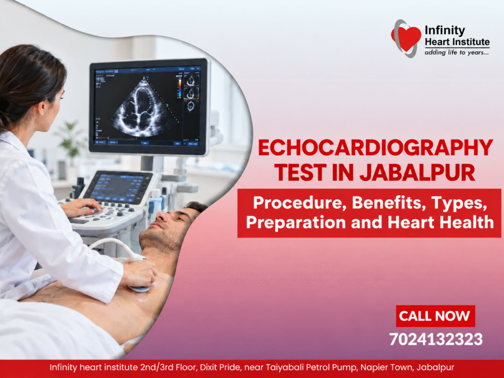
Echocardiography Test in Jabalpur: Procedure, Benefits, Types, Preparation and Heart Health
An Echocardiography Test in Jabalpur is an important diagnostic investigation used to assess the structure and function of the heart. Commonly called an echocardiogram or 2D echo, this test uses ultrasound waves to create moving images of the heart. It helps doctors examine the heart chambers, valves, muscle movement and blood flow without using ionising radiation. Doctors may recommend an echocardiography test when a person experiences breathlessness, chest discomfort, unexplained fatigue, palpitations or symptoms that may indicate a heart condition. It can also help monitor previously diagnosed heart disease and assess cardiac function over time. Patients researching an Echocardiography Specialist in Jabalpur should check the clinician’s qualifications, the diagnostic facilities available and how the report will be interpreted. The appropriate investigation depends on medical history, symptoms and clinical examination. What Is an Echocardiography Test? An echocardiography test is a non-invasive investigation that uses high-frequency sound waves to produce moving images of the heart. A standard transthoracic echocardiogram involves placing an ultrasound probe on the chest to capture images from different angles. The examination helps doctors assess: The size and shape of the heart chambers The thickness and movement of the heart muscle The opening and closing of heart valves The heart’s pumping function Blood flow through the heart Certain structural abnormalities Fluid accumulation around the heart An Echocardiography Test in Jabalpur may be recommended when clinical findings suggest that an assessment of heart structure or function is necessary. It is important to understand that an echocardiogram does not replace every other cardiac investigation. An ECG records electrical activity, whereas an echocardiogram provides images of the heart’s structure and movement. Patients considering a 2D echo should confirm that the examination matches their doctor’s referral and that a qualified professional will review the findings. Why Is Echocardiography Important for Heart Health? The heart pumps blood throughout the body, delivering oxygen and nutrients to organs and tissues. Problems involving the heart muscle, valves or blood flow can interfere with this process. Echocardiography helps doctors investigate these changes and decide whether further evaluation, treatment or follow-up is required. An Echocardiography Test in Jabalpur may be used to assess suspected heart failure, valve disease, enlarged heart chambers, reduced pumping function, congenital heart abnormalities, fluid around the heart and selected complications after a heart attack. A 2D echo may be considered when the clinician needs to examine valve movement, chamber dimensions or pumping function. Not every episode of fatigue or chest discomfort requires an echocardiogram; the decision should be based on clinical need. What Conditions Can an Echocardiogram Detect? 1. Heart Valve Disease Heart valves control the direction of blood flow through the heart. If a valve becomes narrowed or does not close properly, blood flow may be affected. Echocardiography helps assess valve movement, structure and the severity of many valve abnormalities. 2. Reduced Pumping Function An echocardiogram can help evaluate how effectively the heart pumps blood. One measurement commonly included in the report is the left ventricular ejection fraction, which represents the percentage of blood pumped out of the left ventricle with each contraction. 3. Heart Chamber Enlargement An echocardiogram can show whether one or more heart chambers are enlarged. This may occur with valve disorders, cardiomyopathy, congenital conditions or other cardiac problems. 4. Cardiomyopathy Cardiomyopathy refers to diseases affecting the heart muscle. Echocardiography helps assess changes in muscle thickness, chamber size and pumping function. 5. Fluid Around the Heart Fluid can accumulate in the sac surrounding the heart, a condition known as pericardial effusion. Echocardiography can identify the fluid and help doctors assess whether it affects cardiac function. 6. Congenital Heart Conditions Some structural abnormalities present from birth can be detected or evaluated using echocardiography. An echocardiogram may be recommended when a clinician needs to assess heart anatomy, valve function or pumping performance. What Is a 2D Echo Test? A 2D echo test produces two-dimensional moving images of the heart in real time. It allows clinicians to observe the chambers, valves and heart muscle from different angles. Doppler techniques may be used alongside 2D imaging to assess the direction and speed of blood flow. A Heart Ultrasound Test in Jabalpur generally refers to ultrasound-based cardiac imaging. The precise examination depends on the clinical question and the type of echocardiography requested. Patients arranging a 2D echo should confirm the type of scan available, whether the report is included and how the findings will be reviewed. The report should be interpreted by an appropriately qualified medical professional. How Is an Echocardiography Test Performed? A standard transthoracic echocardiogram is usually straightforward and does not require an incision. Step 1: Preparation The patient may be asked to remove clothing from the upper body and wear a medical gown. Small electrodes may be attached to the chest to monitor heart rhythm. Step 2: Positioning The patient generally lies on an examination bed, often turned slightly onto the left side to help obtain clearer images. Step 3: Applying Ultrasound Gel A small amount of gel is applied to the chest to improve contact between the ultrasound probe and the skin. Step 4: Capturing Images The clinician or trained sonographer moves the probe across different areas of the chest. The patient may be asked to change position or briefly adjust breathing. Step 5: Reviewing the Findings The images and measurements are reviewed and interpreted by an appropriately qualified clinician. The findings are considered alongside symptoms, medical history and other investigations. A routine echocardiogram is generally painless, although mild pressure from the probe may be felt. Patients can ask the medical team about the expected duration and when the report will be available. How Long Does an Echocardiogram Take? A standard transthoracic echocardiogram commonly takes around 30–60 minutes. The duration may vary according to the complexity of the examination and the images required. A Cardiac Imaging Test in Jabalpur may involve echocardiography or another investigation, depending on the doctor’s recommendation. Patients should confirm which investigation has been scheduled and check the appointment details before attending. Different Types of Echocardiography Transthoracic Echocardiography